The cranial cruciate ligament (CCL) is an important ligament inside the knee (stifle) joints of dogs. In people it is referred to as the anterior cruciate ligament (ACL). The ligament plays an important role in stabilizing the stifle during weight-bearing. It prevents the shin bone (tibia) moving forwards relative to the thigh bone (femur).In dogs the cruciate ligament tends to undergo degenerative changes that weaken it prior to rupturing. This is in contrast to people, where rupture is often associated with an injury such as skiing or playing football. This important difference explains why treatment options in dogs are quite different to those in people.The reason the cruciate ligament degenerates prior to rupturing is not clearly understood. Certain breeds, such as Labradors and Rottweilers, are much more commonly affected than others. This suggests there is an inherited component to the condition related to conformation of bones or gait.
Rupture of the CCL is the most common cause of hind limb lameness in dogs. Large and giant breeds are particularly affected although any breed and size of dog can rupture their CCL. Some dogs can be affected before they are two years of age.
Is the CCL rupture associated with any other stifle problems?
Rupture of the CCL is associated with the development of osteoarthritis within the stifle. This occurs in every case and is often evident by the time the dog is examined and radiographed. Osteoarthritis tends to be a progressive disorder and treatment of the CCL rupture can reduce or stop this progression.
Many dogs with CCL rupture tear their cartilages (or menisci) within their knees. This is similar to the situation in people. Torn cartilages can be very painful. The response to painkillers can often be poor. Surgery to remove the damaged section of cartilage is generally necessary. Occasionally rupture of the CCL is associated with an abnormality of the shin bone (tibia). It is thought that the bone grows abnormally and this places abnormal forces on the ligament. The ligament subsequently degenerates, weakens and ruptures.
How is CCL Rupture Diagnosed?
The signs of CCL rupture can be quite variable as rupture may be sudden and complete, or gradual and partial. The key signs are hind limb lameness and stiffness. The latter is generally most evident after rest following exercise. Difficulty rising and jumping are common features in dogs with both knees affected. Occasionally ‘clicking’ noises may be heard.Examination may reveal muscle wastage (atrophy), especially over the front of the thigh (the quadriceps muscles). Thickening of the stifle is often palpable. Manipulation of the joint may enable the detection of instability, especially with a cranial drawer. Flexion and extension of the joint may cause pain. In some cases palpation under sedation or light anaesthesia may be necessary to enable the detection of more subtle instability of the knee as occurs with partial rupture of the CCL.
Radiographs provide additional information, especially regarding the presence and severity of osteoarthritis. Specific views may be necessary to assess the angle of the top of the shin bone (the tibial plateau) prior to possible surgery which assists in choosing the correct surgical technique.
Treatment
Some dogs with CCL rupture can be managed satisfactorily without the need for surgery. The smaller the dog, the more likely it is that this approach will be successful. Exercise needs to be restricted. Hydrotherapy is often beneficial. Dogs that are overweight benefit from being placed on a diet. Regular monitoring of weight may be necessary. Painkillers (anti-inflammatory drugs) may be indicated to make the dog more comfortable.
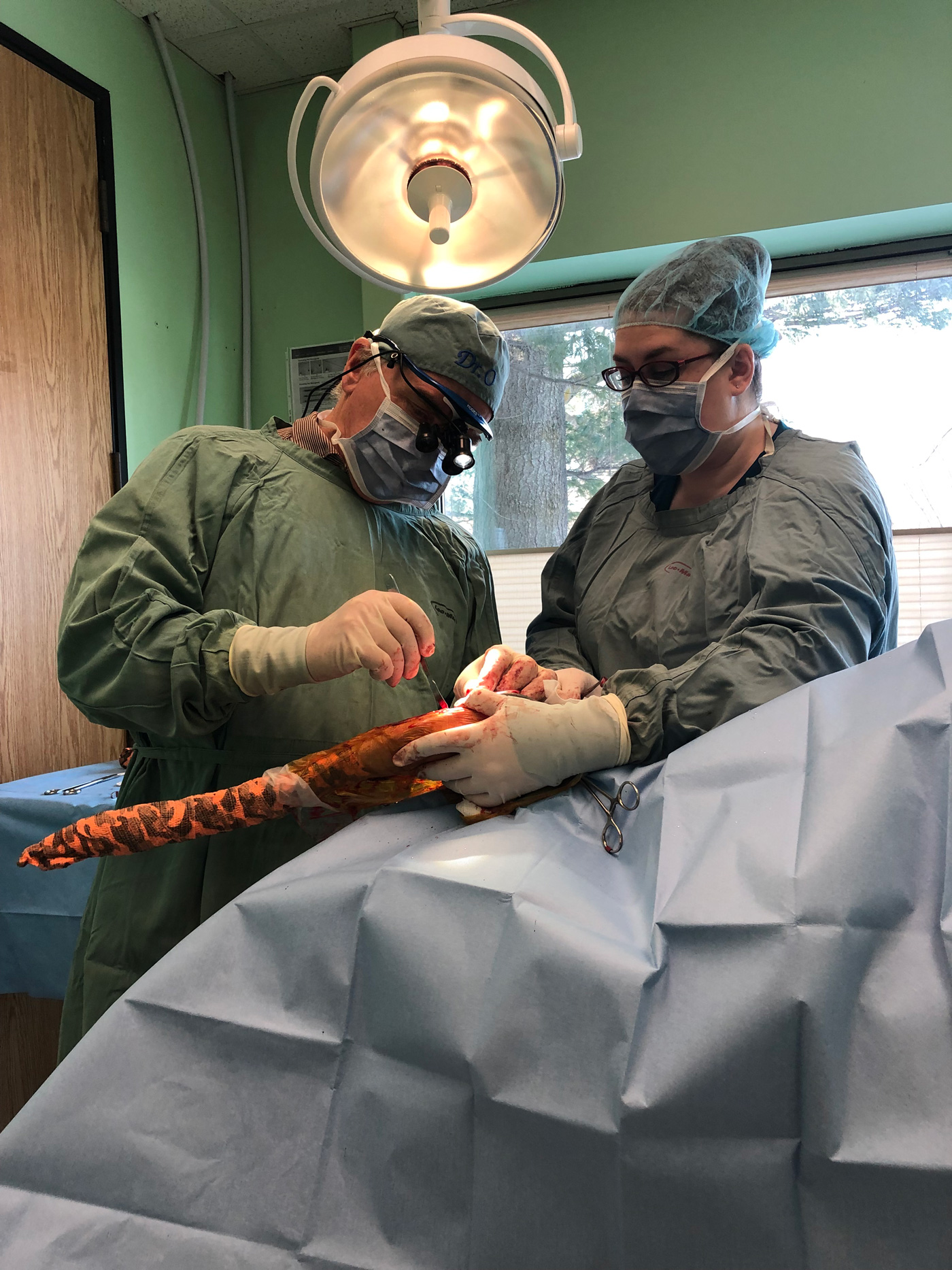
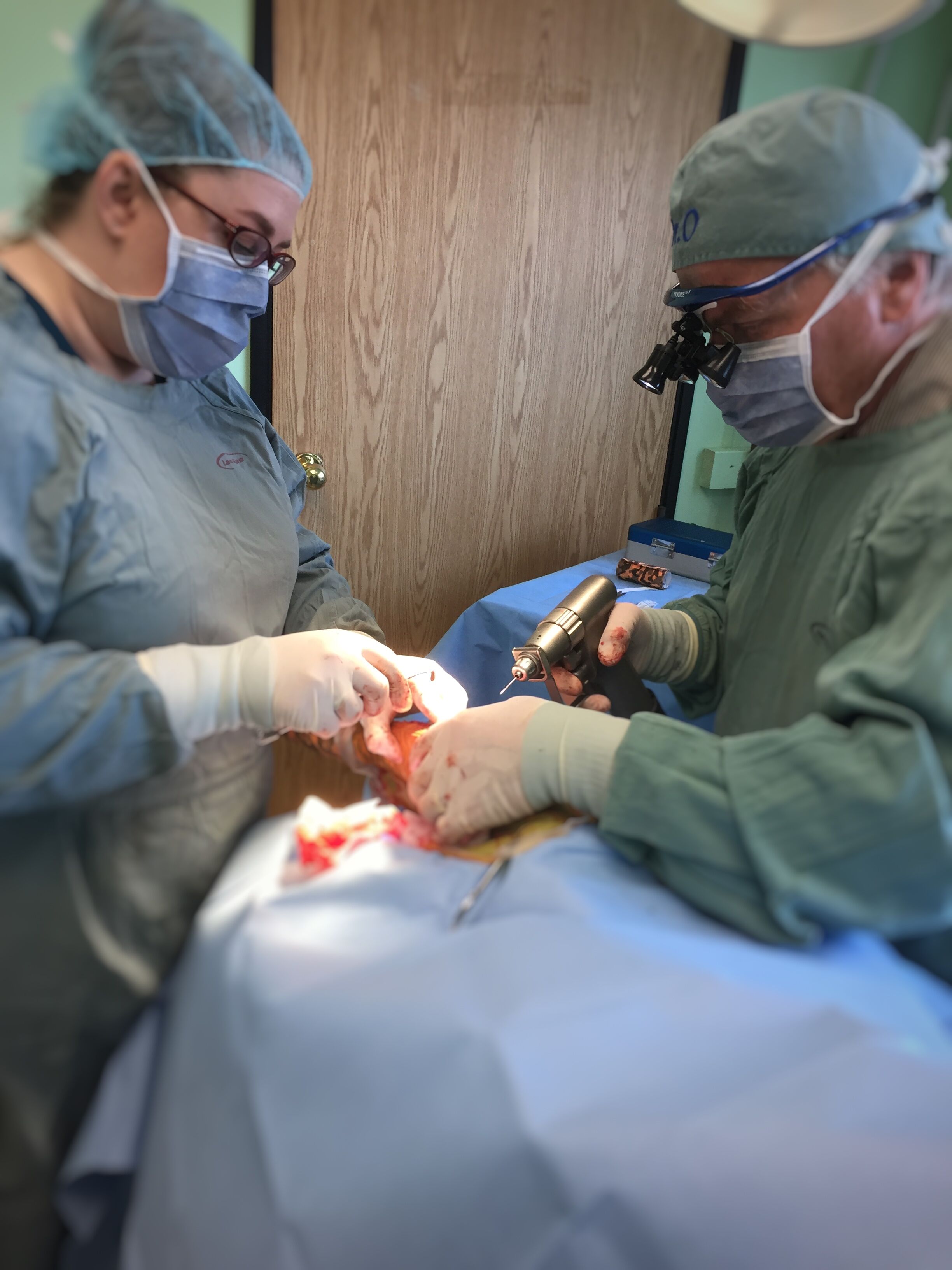
Many medium, large and giant breed dogs with CCL rupture benefit from surgery. There are many types of surgeries that can help. Choosing the best technique depends on the breed, weight, tibial plateau angle, and bone conformation. At our hospitals, Dr. A. Olender is our primary orthopedic surgery. We have the capability of using the TTA technique, meniscal release, and various forms of extracapsular stabilization. For dogs that would benefit most from the TPLO (Tibial Plateau Leveling Osteotomy) technique, they are often referred to a boarded veterinary surgeon. Each procedure holds a good prognosis for stabilizing the joint and return to function, although an increased risk of osteoarthritis is expected in comparison with dogs with intact CCLs.
Extracapsular repair like the Arthrex TightRope, SwivelLock Knottless, Corkscrew and FASTak techniques are all performed our hospitals. These methods use a customized needle and a special suture material affixed to bone anchors or buttons. Each is a variant of the original TightRope procedure. The TightRope technique requires drilling two bone channels, one from side to side through the tibia and the other from side to side through the femur, to run the suture material through which stabilizes the joint. The use of bone anchors help reduce the need for additional suture material in the joint.
TTA is the abbreviation for tibial tuberosity advancement. The procedure involves cutting the front of the tibia, moving it forward, and stabilizing it in a new position with a plate and screws. The surgery aims to make the tibial plateau perpendicular to the patellar tendon which prevents the shin bone moving forward. The knee then feels stable for the dog when weight-bearing, despite the fact that the ligament has been ruptured and not directly repaired
For additional information, the American College of Veterinary Surgeons has an excellent summary. Click here for more details (no actual website URL listed, but please add a button below that links to the page
https://www.acvs.org/small-animal/cranial-cruciate-ligament-disease


